Term Where Body Part Grows Again
Regeneration in humans is the regrowth of lost tissues or organs in response to injury. This is in contrast to wound healing, or partial regeneration, which involves closing up the injury site with some gradation of scar tissue. Some tissues such as peel, the vas deferens, and large organs including the liver can regrow quite readily, while others have been thought to have little or no capacity for regeneration post-obit an injury.
Numerous tissues and organs take been induced to regenerate. Bladders take been 3D-printed in the lab since 1999. Skin tissue can be regenerated in vivo or in vitro. Other organs and torso parts that have been procured to regenerate include: penis, fats, vagina, brain tissue, thymus, and a scaled downwardly human heart. One goal of scientists is to induce full regeneration in more human organs.
In that location are various techniques that tin induce regeneration. By 2016, regeneration of tissue had been induced and operationalized by scientific discipline. In that location are iv chief techniques: regeneration by instrument;[1] regeneration past materials;[2] [three] regeneration by drugs[4] [5] [vi] and regeneration past in vitro 3D printing.[3]
History of homo tissue [edit]
In humans with non-injured tissues, the tissue naturally regenerates over time; by default, new available cells replace expended cells. For example, the body regenerates a full bone within ten years, while non-injured skin tissue is regenerated within 2 weeks.[2] With injured tissue, the body usually has a different response. This emergency response usually involves building a caste of scar tissue over a time period longer than a regenerative response, as has been proven clinically[7] and via observation. There are many more historical and nuanced understandings well-nigh regeneration processes. In total thickness wounds that are under 2mm, regeneration generally occurs before scarring.[eight] In 2008, in full thickness wounds over 3mm, information technology was establish that a wound needed a cloth inserted in order to induce total tissue regeneration.[nine] [10]
Some human organs and tissues regenerate rather than simply scar, as a effect of injury. These include the liver, fingertips, and endometrium. More information is now known regarding the passive replacement of tissues in the human being body, as well as the mechanics of stem cells. Advances in enquiry have enabled the induced regeneration of many more tissues and organs than previously thought possible. The aim for these techniques is to use these techniques in the near future for the purpose of regenerating any tissue type in the human body.
Regeneration techniques [edit]
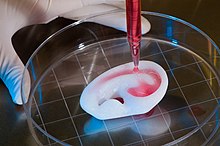
Regenerating a human ear using a scaffold
By 2016, regeneration had been operationalised and induced past 4 main techniques: regeneration by musical instrument;[1] regeneration by materials;[2] [3] regeneration by 3d press;[3] and regeneration past drugs.[4] [5] [6] By 2016, regeneration by instrument, regeneration by materials and by regeneration drugs had been mostly operationalised in vivo (inside living tissues). Whilst by 2016, regeneration by 3d press had been mostly operationalised by in vitro (within the lab) in order to be built and prepare tissue for transplantation.[3]
By musical instrument [edit]
A cut by a pocketknife or a scalpel generally scars, though a piercing past a needle does non.[1] [11] In 1976, a 3 by 3cm scar on a non-diabetic was regenerated by insulin injections and the researchers, highlighting earlier research, argued that the insulin was regenerating the tissue.[4] [5] The anecdotal evidence likewise highlighted that a syringe was one of two variables that helped bring regeneration of the arm scar.[4] The syringe was injected into the iv quadrants iii times a day for eighty-two days.[4] Afterward eighty-two days, afterward many sequent injections, the scar was resolved and it was noted no scar was appreciable past the human heart.[4] After 7 months the area was checked again and it was once more noted that no scar could exist seen.[four]
In 1997, it was proven that wounds created with an instrument that are under 2mm can heal scar complimentary,[8] just larger wounds that are larger than 2mm healed with a scar.[8]
In 2013, information technology was proven in pig tissue that full thickness micro columns of tissue, less than 0.5mm in diameter could be removed and that the replacement tissue, was regenerative tissue, not scar. The tissue was removed in a fractional pattern, with over 40% of a square area removed; and all of the fractional full thickness holes in the foursquare area healed without scarring.[12] In 2016 this fractional blueprint technique was also proven in human tissue.[one]
With materials [edit]
More often than not, humans tin regenerate injured tissues in vivo for limited distances of upwards to 2mm. The further the wound altitude is from 2mm the more the wound regeneration will demand inducement. By 2009, via the utilise of materials, a max induced regeneration could exist achieved inside a i cm tissue rupture.[2] Bridging the wound, the material immune cells to cantankerous the wound gap; the textile then degraded. This engineering was first used inside a broken urethra in 1996.[2] [3] In 2012, using materials, a full urethra was restored in vivo.[3]
Macrophage polarization is a strategy for skin regeneration.[xiii] Macrophages are differentiated from circulating monocytes.[thirteen] Macrophages display a range of phenotypes varying from the M1, pro-inflammatory type to the M2, pro-regenerative type.[13] Cloth hydrogels polarise macrophages into the key M2 regenerative phenotype in vitro.[13] In 2017, hydrogels provided full regeneration of peel, with hair follicles, afterwards partial excision of scars in pigs and subsequently full thickness wound incisions in pigs.[xiii]
By 3D press [edit]
In 2009, the regeneration of hollow organs and tissues with a long diffusion altitude, was a lilliputian more challenging. Therefore, to regenerate hollow organs and tissues with a long diffusion distance, the tissue had to exist regenerated inside the lab, via the use of a 3D printer.[2]
Diverse tissues that have been regenerated past in vitro 3D press include:
- The starting time organ ever induced and made in the lab was the float, which was created in 1999.[14]
- By 2014, there had been various tissues regenerated past the 3D printer and these tissues included: muscle, vagina, penis and the thymus.
- In 2014, a conceptual human lung was first bioengineered in the lab.[15] [xvi] In 2015, the lab robustly tested its technique and regenerated a pig lung.[xv] [16] The pig lung was and so successfully transplanted into a hog without the apply of immunosuppressive drugs.[xv] [sixteen]
- In 2015, researchers developed a proof of principle biolimb inside a laboratory; they also estimated that it would exist at least a decade for any testing of limbs in humans. The limb demonstrated fully functioning skin, muscles, blood vessels and bones.[17]
- In April 2019, researchers 3d printed a homo heart.[18] The image middle was made by human stem cells but just to the size of a rabbit's heart.[18] In 2019, the researchers hoped to one twenty-four hours place a scaled upwardly version of the center within humans.[eighteen]
Gradations of complexity [edit]
| Level 1 | Level 2 | Level iii | Level four |
|---|---|---|---|
| Skin | Blood vessel | Bladder | Heart |
| Muscle | Liver | ||
| Nails | Pancreas | ||
| Penis |
With printing tissues, by 2012, there were four accustomed standard levels of regenerative complexity that were acknowledged in various academic institutions:
- Level i, apartment tissue like skin was the simplest to recreate;[3]
- Level two was tubular structures such every bit blood vessels;[iii]
- Level iii was hollow not-tubular structures;[3]
- Level 4 was solid organs, which were by far the most circuitous to recreate due to the vascularity.[3]
In 2012, within 60 days it was possible, inside the lab, to grow tissue the size of one-half a postage stamp to the size of a football field. Most prison cell types could be grown and expanded exterior of the body, with the exception of the liver, nerve and pancreas, as these tissue types need stem cell populations.[three]
With drugs [edit]
Lipoatrophy is the localised loss of fatty in tissue. Information technology is common in diabetics who use conventional insulin injection treatment.[4] In 1949, a much more than pure grade of insulin was, instead of causing lipoatrophy, shown to regenerate the localised loss of fat after injections in to diabetics.[iv] In 1984, it was shown that dissimilar insulin injections have different regenerative responses with regards to creating skin fats in the aforementioned person.[five] Information technology was shown in the same torso that conventional forms of insulin injections cause lipoatrophy and highly purified insulin injections crusade lipohypertrophy.[v] In 1976, the regenerative response was shown to work in a non-diabetic afterward a iii ten 3cm lipoatrophic arm scar was treated with pure monocomponent porcine soluble insulin.[five] [iv] A syringe injected insulin under the skin equally in the four quadrants of the defect.[4] To layer four units of insulin evenly into the base of the defect, each quadrant of the defect received one unit of insulin three times a day, for fourscore-2 days.[4] After fourscore-2 days of consecutive injections the defect regenerated to normal tissue.[iv] [5]
In 2016, scientists could transform a peel cell into any other tissue type via the use of drugs.[half-dozen] The technique was noted as safer than genetic reprogramming which, in 2016, was a concern medically.[6] The technique, used a cocktail of chemicals and enabled efficient on site regeneration without whatever genetic programming.[6] In 2016, it was hoped to one day apply this drug to regenerate tissue at the site of tissue injury.[vi] In 2017, scientists could plow many jail cell types (such as brain and center) into skin.[19]
Naturally regenerating appendages and organs [edit]
Heart [edit]
Cardiomyocyte necrosis activates an inflammatory response that serves to clear the injured myocardium from dead cells, and stimulates repair, but may also extend injury. Research suggests that the cell types involved in the process play an important office. Namely monocyte-derived macrophages tend to induce inflammation while inhibiting cardiac regeneration, while tissue resident macrophages may help restoration of tissue structure and function.[20]
Endometrium [edit]
The endometrium after the process of breakdown via the menstruation bike, re-epithelializes swiftly and regenerates.[21] Though tissues with a non-interrupted morphology, like non-injured soft tissue, completely regenerate consistently; the endometrium is the merely man tissue that completely regenerates consistently afterwards a disruption and pause of the morphology.[21]
Fingers [edit]
In May 1932, L.H. McKim published a report describing the regeneration of an adult digit-tip following amputation. A house surgeon in the Montreal General Hospital underwent amputation of the distal phalanx to finish the spread of an infection. In less than one month following surgery, x-ray assay showed the regrowth of bone while macroscopic ascertainment showed the regrowth of boom and skin.[22] This is one of the earliest recorded examples of developed human being digit-tip regeneration.[23]
Studies in the 1970s showed that children upward to the age of x or so who lose fingertips in accidents can regrow the tip of the digit within a month provided their wounds are not sealed upward with flaps of peel – the de facto treatment in such emergencies. They normally won't have a fingerprint, and if there is whatever piece of the finger nail left it will grow back as well, usually in a square shape rather than round.[24] [25]
In Baronial 2005, Lee Spievack, then in his early sixties, accidentally sliced off the tip of his right middle finger just above the starting time phalanx. His brother, Dr. Alan Spievack, was researching regeneration and provided him with powdered extracellular matrix, adult past Dr. Stephen Badylak of the McGowan Institute of Regenerative Medicine. Mr. Spievack covered the wound with the powder, and the tip of his finger re-grew in four weeks.[26] The news was released in 2007. Ben Goldacre has described this every bit "the missing finger that never was", claiming that fingertips regrow and quoted Simon Kay, professor of mitt surgery at the Academy of Leeds, who from the pic provided past Goldacre described the case equally seemingly "an ordinary fingertip injury with quite unremarkable healing"[27]
A similar story was reported past CNN. A woman named Deepa Kulkarni lost the tip of her little finger and was initially told by doctors that nothing could be done. Her personal inquiry and consultation with several specialists including Badylak eventually resulted in her undergoing regenerative therapy and regaining her fingertip.[28]
Kidney [edit]
Regenerative chapters of the kidney has been recently explored.[29]
The basic functional and structural unit of the kidney is nephron, which is mainly composed of four components: the glomerulus, tubules, the collecting duct and peritubular capillaries. The regenerative capacity of the mammalian kidney is express compared to that of lower vertebrates.
In the mammalian kidney, the regeneration of the tubular component following an acute injury is well known. Recently regeneration of the glomerulus has also been documented. Following an acute injury, the proximal tubule is damaged more, and the injured epithelial cells slough off the basement membrane of the nephron. The surviving epithelial cells, however, undergo migration, dedifferentiation, proliferation, and redifferentiation to furnish the epithelial lining of the proximal tubule afterward injury. Recently, the presence and participation of kidney stem cells in the tubular regeneration has been shown. However, the concept of kidney stem cells is currently emerging. In addition to the surviving tubular epithelial cells and kidney stalk cells, the os marrow stem cells have as well been shown to participate in regeneration of the proximal tubule, however, the mechanisms remain controversial. Studies examining the capacity of bone marrow stem cells to differentiate into renal cells are emerging.[thirty]
Like other organs, the kidney is too known to regenerate completely in lower vertebrates such as fish. Some of the known fish that show remarkable capacity of kidney regeneration are goldfish, skates, rays, and sharks. In these fish, the entire nephron regenerates post-obit injury or partial removal of the kidney.
Liver [edit]
The human being liver is particularly known for its ability to regenerate, and is capable of doing then from only i quarter of its tissue,[31] due chiefly to the unipotency of hepatocytes.[32] Resection of liver can induce the proliferation of the remaining hepatocytes until the lost mass is restored, where the intensity of the liver'south response is directly proportional to the mass resected. For almost 80 years surgical resection of the liver in rodents has been a very useful model to the study of jail cell proliferation.[33] [34]
Toes [edit]
Toes damaged by gangrene and burns in older people can also regrow with the blast and toe impress returning afterwards medical treatment for gangrene.[35]
Vas deferens [edit]
The vas deferens can abound dorsum together after a vasectomy–thus resulting in vasectomy failure.[36] This occurs due to the fact that the epithelium of the vas deferens, similar to the epithelium of some other human body parts, is capable of regenerating and creating a new tube in the upshot that the vas deferens is damaged and/or severed.[37] Even when as much as v centimeters, or two inches, of the vas deferens is removed, the vas deferens can withal grow back together and get reattached–thus assuasive sperm to once once again pass and flow through the vas deferens, restoring one's fertility.[37]
Induced regeneration [edit]
There are several human tissues that take been successfully or partially induced to regenerate. Many fall under the topic of regenerative medicine, which includes the methods and inquiry conducted with the aim of regenerating the organs and tissues of humans as a consequence of injury. The major strategies of regenerative medicine include dedifferentiating injury site cells, transplanting stem cells, implanting lab-grown tissues and organs, and implanting bioartificial tissues.
Bladder [edit]
In 1999, the float was the first regenerated organ to be given to seven patients; equally of 2014, these regenerated bladders are still functioning inside the beneficiaries.[14]
Fatty [edit]
In 1949, purified insulin was shown to regenerate fat in diabetics with lipoatrophy.[4] In 1976, later 82 days of consecutive injections into a scar, purified insulin was shown to safely regenerate fat and completely regenerate skin in a non-diabetic.[4] [5]
During a loftier-fat diet, and during pilus follicle growth, mature adipocytes (fats) are naturally formed in multiple tissues.[38] Fat tissue has been implicated in the inducement of tissue regeneration. Myofibroblasts are the fibroblast responsible for scar and in 2017 it was found that the regeneration of fat transformed myofibroblasts into adipocytes instead of scar tissue.[39] [38] Scientists also identified bone morphogenetic protein (BMP) signalling equally important for myofibroblasts transforming into adipocytes for the purpose of skin and fat regeneration.[39]
Heart [edit]
Cardiovascular diseases are the leading crusade of death worldwide, and accept increased proportionally from 25.8% of global deaths in 1990, to 31.5% of deaths in 2013.[twoscore] This is true in all areas of the world except Africa.[twoscore] [41] In add-on, during a typical myocardial infarction or center attack, an estimated one billion cardiac cells are lost.[42] The scarring that results is then responsible for profoundly increasing the take a chance of life-threatening abnormal heart rhythms or arrhythmias. Therefore, the power to naturally regenerate the eye would have an enormous impact on modern healthcare. Notwithstanding, while several animals tin regenerate center harm (e.yard. the axolotl), mammalian cardiomyocytes (middle muscle cells) cannot proliferate (multiply) and centre damage causes scarring and fibrosis.
Despite the earlier belief that human cardiomyocytes are non generated later on in life, a recent study has found that this is not the case. This study took reward of the nuclear bomb testing during the Common cold War, which introduced carbon-14 into the atmosphere and therefore into the cells of nearby inhabitants.[43] They extracted Dna from the myocardium of these research subjects and found that cardiomyocytes do in fact renew at a slowing charge per unit of 1% per year from the age of 25, to 0.45% per year at the historic period of 75.[43] This amounts to less than half of the original cardiomyocytes being replaced during the average lifespan. All the same, serious doubts have been placed on the validity of this enquiry, including the ceremoniousness of the samples every bit representative of normally aging hearts.[44]
Farther research has been conducted that supports the potential for human cardiac regeneration. Inhibition of p38 MAP kinase was establish to induce mitosis in adult mammalian cardiomyocytes,[45] while treatment with FGF1 and p38 MAP kinase inhibitors was found to regenerate the heart, reduce scarring, and improve cardiac part in rats with cardiac injury.[46]
I of the most promising sources of heart regeneration is the utilise of stem cells. It was demonstrated in mice that there is a resident population of stem cells or cardiac progenitors in the adult heart – this population of stem cells was shown to be reprogrammed to differentiate into cardiomyocytes that replaced those lost during a heart tissue death.[47] In humans specifically, a "cardiac mesenchymal feeder layer" was found in the myocardium that renewed the cells with progenitors that differentiated into mature cardiac cells.[48] What these studies show is that the human being center contains stem cells that could potentially exist induced into regenerating the center when needed, rather than but being used to supplant expended cells.
Loss of the myocardium due to disease often leads to heart failure; therefore, it would be useful to be able to take cells from elsewhere in the center to replenish those lost. This was accomplished in 2010 when mature cardiac fibroblasts were reprogrammed directly into cardiomyocyte-similar cells. This was done using 3 transcription factors: GATA4, Mef2c, and Tbx5.[49] Cardiac fibroblasts make up more than half of all middle cells and are usually non able to conduct contractions (are not cardiogenic), but those reprogrammed were able to contract spontaneously.[49] The significance is that fibroblasts from the damaged centre or from elsewhere, may be a source of functional cardiomyocytes for regeneration.
But injecting functioning cardiac cells into a damaged center is only partially effective. In order to reach more reliable results, structures equanimous of the cells demand to be produced and then transplanted. Masumoto and his team designed a method of producing sheets of cardiomyocytes and vascular cells from human iPSCs. These sheets were then transplanted onto infarcted hearts of rats, leading to significantly improved cardiac function.[50] These sheets were still establish to be present four weeks later.[l] Research has too been conducted into the engineering of heart valves. Tissue-engineered heart valves derived from human cells have been created in vitro and transplanted into a non-man primate model. These showed a promising amount of cellular repopulation even afterwards eight weeks, and succeeded in outperforming currently-used not-biological valves.[51] In 2021, researchers demonstrated a switchable iPSCs-reprogramming-based arroyo for regeneration of damaged middle without tumor-formation in mice.[52] In April 2019, researchers 3d printed a prototype human heart the size of a rabbit'due south heart.[xviii]
Lung [edit]
Chronic obstructive pulmonary disease (COPD) is one of the near widespread health threats today. It affects 329 million people worldwide, which makes up virtually 5% of the global population. Having killed over 3 million people in 2012, COPD was the third greatest cause of decease.[53] Worse still, due to increasing smoking rates and the aging populations in many countries, the number of deaths as a result of COPD and other chronic lung diseases is predicted to continue increasing.[54] Therefore, developments in the lung's capacity for regeneration is in high demand.
It has been shown that bone marrow-derived cells could be the source of progenitor cells of multiple cell lineages, and a 2004 written report suggested that one of these cell types was involved in lung regeneration.[55] Therefore, a potential source of cells for lung regeneration has been constitute; however, due to advances in inducing stalk cells and directing their differentiation, major progress in lung regeneration has consistently featured the use of patient-derived iPSCs and bioscaffolds. The extracellular matrix is the central to generating entire organs in vitro. It was found that past carefully removing the cells of an entire lung, a "footprint" is left behind that can guide cellular adhesion and differentiation if a population of lung epithelial cells and chondrocytes are added.[56] This has serious applications in regenerative medicine, peculiarly as a 2012 written report successfully purified a population of lung progenitor cells that were derived from embryonic stem cells. These tin can then be used to re-cellularise a three-dimensional lung tissue scaffold.[57]
Indeed, in 2008, there was a successful clinical transplantation of a tissue-engineered trachea in a 30-year-old adult female with end-stage bronchomalacia. An ECM scaffold was created by removing the cells and MHC antigens from a human donated trachea, which was then colonised by epithelial cells and mesenchymal stem prison cell-derived chondrocytes cultured from cells of the recipient.[58] The graft replaced her left main bronchus, immediately providing a functional airway, and retained its normal appearance and mechanical part after four months.[58] Because the graft was generated from cells cultured from the recipient, no anti-donor antibodies or immunosuppressive drugs were needed—a huge step towards personalised lung regeneration.
A 2010 investigation took this one pace further past using the ECM scaffold to produce entire lungs in vitro to be transplanted into living rats.[59] These successfully enabled gas substitution but for brusque time intervals only.[59] Nevertheless, this was a huge leap towards whole lung regeneration and transplants for humans, which has already taken another step forward with the lung regeneration of a not-human primate.[60]
Cystic fibrosis is another disease of the lungs, which is highly fatal and genetically linked to a mutation in the CFTR factor. Through growing patient-specific lung epithelium in vitro, lung tissue expressing the cystic fibrosis phenotype has been achieved.[61] This is and then that modelling and drug testing of the illness pathology can be carried out with the hope of regenerative medical applications.
Penis [edit]
Penises have been successfully regenerated in the lab.[14] Penises are harder to regenerate than the skin, bladder and vagina due to their structural complexity.[fourteen]
Spinal nerves [edit]
A goal of spinal cord injury research is to promote neuroregeneration, reconnection of damaged neural circuits.[62] The fretfulness in the spine are a tissue that requires a stem cell population to regenerate. In 2012, a Polish fireman Darek Fidyka, with paraplegia of the spinal cord, underwent a process, which involved extracting olfactory ensheathing cells (OECs) from Fidyka's olfactory bulbs, and injecting these stem cells, in vivo, into the site of the previous injury. Fidyka somewhen gained feeling, move and sensation in his limbs, especially on the side where the stalk cells were injected; he besides reported gaining sexual role. Fidyka tin can now drive and can now walk some distance aided by a frame. He is believed to be the beginning person in the earth to recover sensory function from a consummate severing of the spinal nerves.[63] [64]
Thymus [edit]
The thymus gland is 1 of the first organs to degenerate in normal healthy individuals. Researchers from the University of Edinburgh have succeeded in regenerating a living organ that closely resembles a juvenile thymus in terms of structure and gene expression contour.[65]
Vagina [edit]
Between the years 2005 and 2008, 4 women with vaginal hypoplasia due to Müllerian agenesis were given regenerated vaginas.[66] Upwardly to 8 years subsequently the transplants, all organs take normal function and structure.[14]
See also [edit]
- Tissue engineering
References [edit]
- ^ a b c d Tam, Joshua (14 June 2016). "Reconstitution of total-thickness skin by microcolumn grafting". Journal of Tissue Technology and Regenerative Medicine. 11 (10): 2796–2805. doi:10.1002/term.2174. PMC5697650. PMID 27296503.
- ^ a b c d e f Atala, Anthony (October 2009). "Growing new organs". TED.
- ^ a b c d due east f k h i j k fifty McManus, Rich (2 March 2012). "Atala Surveys Successes of Regenerative Medicine". nihrecord.nih.gov. Archived from the original on 2014-11-21. Retrieved 7 April 2015.
- ^ a b c d eastward f g h i j grand l grand n o Amroliwalla, F Grand. (25 March 1977). "Vaccination scar with soft-tissue atrophy restored by local insulin treatment". British Medical Journal. 1 (6073): 1389–1390. doi:ten.1136/bmj.1.6073.1389. PMC1606939. PMID 861647.
- ^ a b c d e f g h Campbell, W; Duncan, C; Anani, A. R. (1984). "Paradoxical lipodystrophic changes due to conventional bovine and highly purified porcine/bovine insulins". British Medical Journal. Britain: pmj.bmj.com. 60 (704): 439–441. doi:10.1136/pgmj.60.704.439. PMC2417884. PMID 6379631.
- ^ a b c d e f Smith, Dana Grand. (28 April 2016). "Scientists plow skin cells into heart cells and brain cells using drugs: Studies represent beginning purely chemical cellular reprogramming, changing a jail cell'southward identity without adding external genes". sciencedaily.com. Gladstone Institutes.
- ^ Cubison TC, Pape SA, Parkhouse Due north (December 2006). "Evidence for the link betwixt healing fourth dimension and the development of hypertrophic scars (HTS) in paediatric burns due to scald injury". Burns. 32 (8): 992–9. doi:10.1016/j.burns.2006.02.007. PMID 16901651.
- ^ a b c Wilgus, Traci A. (June 2007). "Regenerative Healing in Fetal Peel: A Review of the Literature". Ostomy Wound Direction. 53 (6): 16–31, quiz 32-three. PMID 17586870.
- ^ Dorin RP, Pohl HG, De Filippo RE, Yoo JJ, Atala A (2008). "A. World J. Urol. 2008; 26:323". World J Urol. 26 (four): 323–6. doi:10.1007/s00345-008-0316-vi. PMID 18682960. S2CID 24808282.
- ^ Anthony Atala; Darrell J. Irvine; Marsha Moses; Sunil Shaunak (ane August 2010). "Wound Healing Versus Regeneration: Part of the Tissue Environs in Regenerative Medicine". MRS Balderdash. 35 (8): 597–606. doi:10.1557/mrs2010.528. PMC3826556. PMID 24241586.
- ^ Joshua Tam (2013). "Fractional Skin Harvesting: Autologous Skin Grafting without Donor-site Morbidity". Plastic and Reconstructive Surgery. Global Open. Plastic & Reconstructive Surgery September 2013. 1 (half-dozen): e47. doi:x.1097/GOX.0b013e3182a85a36. PMC4174164. PMID 25289241.
- ^ Justin R. Fernandes, MD, Juan C. Samayoa, MD, Thou. Felix Broelsch, MD, Michael C. McCormack, MBA, Alexa M. Nicholls, BS, Mark A. Randolph, MAS, Martin C. Mihm, MD, William G. Austen, Jr., Dr. (2013). "Micro-Mechanical Partial Peel Rejuvenation". Plastic and Reconstructive Surgery. PLASTIC SURGERY 2012. 131 (2): 216–23. doi:10.1097/PRS.0b013e3182789afa. PMID 23357983. S2CID 205973125.
{{cite journal}}: CS1 maint: multiple names: authors list (link) - ^ a b c d east Savoji, Houman; Godau, Brent; Sheikh Hassani, Mohsen; Akbari, Mohsen (26 July 2018). "Peel Tissue Substitutes and Biomaterial Take a chance Cess and Testing". Frontiers in Bioengineering and Biotechnology. Front end. Bioeng. Biotechnol. six: 86. doi:10.3389/fbioe.2018.00086. PMC6070628. PMID 30094235.
- ^ a b c d e Mohammadi, Dara (four Oct 2014). "Bioengineered organs: The story so far…". theguardian.com. Retrieved 9 March 2015.
- ^ a b c Gonzalez, Robbie (8 January 2018). "Bioengineers Are Closer Than Always To Lab-Grown Lungs". wired.com . Retrieved 27 May 2020.
- ^ a b c Uriarte, Juan J (2018). "Lung bioengineering advances and challenges in lung decellularization and recellularization". Current Opinion in Organ Transplantation. journals.lww.com. 23 (6): 673–678. doi:10.1097/MOT.0000000000000584. PMC8669574. PMID 30300330. S2CID 52946782.
- ^ Plaugic, Lizzie (4 June 2015). "Researchers have grown a partially functioning rat limb in a lab". theverge.com. washingtonpost.com. Retrieved viii June 2015.
- ^ a b c d Bracho-Sanchez, Dr. Edith (17 Apr 2019). "Researchers 3D-print heart from human patient's cells". edition.cnn.com. cnn. Retrieved viii May 2019.
- ^ ScienceDaily Staff (18 October 2017). "Turning encephalon cells into peel cells: Researchers transform mature cells from the brain, heart and more into pare cells". sciencedaily.com. American Friends of Tel Aviv University.
- ^ Frangogiannis, Northward.Chiliad. (May 2015). "Inflammation in cardiac injury, repair and regeneration". Curr Opin Cardiol. thirty (3): 240–245. doi:10.1097/HCO.0000000000000158. PMC4401066. PMID 25807226.
- ^ a b Min, Su; Wang, Song West.; Orr, William (2006). "Graphic general pathology: ii.2 complete regeneration". Pathology. pathol.med.stu.edu.cn. Archived from the original on 2012-12-07. Retrieved 2013-11-ten .
Later the repair procedure has been completed, the structure and function of the injured tissue are completely normal. This blazon of regeneration is mutual in physiological situations. Examples of physiological regeneration are the continual replacement of cells of the skin and repair of the endometrium later menstruation. Complete regeneration can occur in pathological situations in tissues that take good regenerative capacity.
- ^ McKim, L.H. (May 1932). "Regeneration Of The Distal Phalanx". The Canadian Medical Clan Journal. 26 (5): 549–550. PMC402335. PMID 20318716.
- ^ Wicker, Hashemite kingdom of jordan; Kenneth Kamler (August 2009). "Current concepts in limb regeneration: A hand surgeon's perspective". Annals of the New York University of Sciences. 1172 (1): 95–109. Bibcode:2009NYASA1172...95W. doi:10.1111/j.1749-6632.2009.04413.x. PMID 19735243. S2CID 22948936.
- ^ Weintraub, Arlene (May 24, 2004). "The Geniuses Of Regeneration". BusinessWeek.
- ^ Illingworth Cynthia G (1974). "Trapped fingers and amputated fingertips in children". Journal of Pediatric Surgery. 9 (six): 853–858. doi:10.1016/s0022-3468(74)80220-4. PMID 4473530.
- ^ "Regeneration recipe: Pinch of pig, cell of lizard". NBC News. Associated Press. February 19, 2007. Retrieved October 24, 2008.
- ^ Goldacre, Ben (May 3, 2008). "The missing finger that never was". The Guardian.
- ^ Adult female's persistence pays off in regenerated fingertip past Elizabeth Cohen. CNN website, September nine, 2010 4:51 p.m., page found 2010-09-xvi.
- ^ Song Jeremy J (2013). "Regeneration and experimental orthotopic transplantation of a bioengineered kidney". Nature Medicine. nineteen (5): 646–651. doi:ten.1038/nm.3154. PMC3650107. PMID 23584091.
- ^ Kurinji; et al. (2009). "In Vitro Differentiation of MSC into Cells with a Renal Tubular Epithelial-Like Phenotype". Renal Failure. 31 (vi): 492–502. doi:10.1080/08860220902928981. PMID 19839827.
- ^ "Liver Regeneration Unplugged". Bio-Medicine. 2007-04-17. Retrieved 2007-04-17 .
- ^ Michael, Sandra Rose (2007). "Bio-Scalar Technology: Regeneration and Optimization of the Body-Mind Homeostasis" (PDF). 15th Almanac AAAAM Conference: 2. Archived from the original (PDF) on November 20, 2008. Retrieved Oct 24, 2008.
- ^ Higgins, GM; RM Anderson RM (1931). "Experimental pathology of the liver. I. Restoration of the liver of the white rat following partial surgical removal". Arch. Pathol. 12: 186–202.
- ^ Michalopoulos, GK; MC DeFrances (Apr 4, 1997). "Liver regeneration". Science. 276 (5309): sixty–66. doi:10.1126/science.276.5309.sixty. PMID 9082986. S2CID 2756510.
- ^ DeMarco, Peter. 1986. Method of treatment of animal and homo tissues damaged by burns and frank visible gangrene. US 4618490
- ^ Miller, Korin (2017-09-11). "Hither'south What Happens When a Vasectomy Fails". SELF . Retrieved 2019-03-sixteen .
- ^ a b Rolnick, H. C. (July 1924). "Regeneration of the Vas Deferens". Athenaeum of Surgery. nine (1): 188. doi:10.1001/archsurg.1924.01120070191008. ISSN 0004-0010.
- ^ a b Horsley, Watt (half-dozen Apr 2017). "Repeal and Replace: Adipocyte Regeneration in Wound Repair". Cell Stalk Cell (Submitted manuscript). 20 (4): 424–426. doi:ten.1016/j.stem.2017.03.015. PMID 28388424.
- ^ a b Plikus; et al. (5 January 2017). "Regeneration of fatty cells from myofibroblasts during wound healing". Science. 355 (6326): 748–752. Bibcode:2017Sci...355..748P. doi:10.1126/science.aai8792. PMC5464786. PMID 28059714.
- ^ a b Mendis, Shanthi; Puska, Pekka; Norrving, Bo (2011). Global atlas on cardiovascular disease prevention and command (PDF) (1st ed.). Geneva: Earth Health Organization in collaboration with the World Heart Federation and the World Stroke Organization. pp. 3–18. ISBN9789241564373.
- ^ GBD 2013 Mortality and Causes of Expiry, Collaborators (17 December 2014). "Global, regional, and national age-sex specific all-cause and crusade-specific bloodshed for 240 causes of death, 1990-2013: a systematic assay for the Global Burden of Affliction Study 2013". Lancet. 385 (9963): 117–71. doi:10.1016/S0140-6736(14)61682-2. PMC4340604. PMID 25530442.
- ^ Laflamme, MA; Murry, CE (July 2005). "Regenerating the heart". Nature Biotechnology. 23 (7): 845–56. doi:10.1038/nbt1117. PMID 16003373. S2CID 8265954.
- ^ a b Bergmann O, et al. (2009). "Evidence for Cariomyocyte Renewal in Humans". Science. 324 (5923): 98–102. Bibcode:2009Sci...324...98B. doi:ten.1126/scientific discipline.1164680. PMC2991140. PMID 19342590.
- ^ Kajstura J, et al. (2012). "Response to Bergmann et al: Carbon fourteen Birth Dating of Human Cardiomyocytes". Circulation Research. 110 (1): e19–e21. doi:10.1161/CIRCRESAHA.111.259721. PMC4159170. PMID 25214670.
- ^ Engel, F. B.; Schebesta, M.; Duong, M. T.; Lu, G.; Ren, S.; Madwed, J. B.; Jiang, H.; Wang, Y.; Keating, M. T. (2005). "P38 MAP kinase inhibition enables proliferation of adult mammalian cardiomyocytes". Genes & Evolution. xix (10): 1175–1187. doi:x.1101/gad.1306705. PMC1132004. PMID 15870258.
- ^ Felix B. Engel, Patrick C. H. Hsieh, Richard T. Lee, Mark T. Keating; Hsieh; Lee; Keating (October 2006). "FGF1/p38 MAP kinase inhibitor therapy induces cardiomyocyte mitosis, reduces scarring, and rescues role later on myocardial infarction". Proceedings of the National University of Sciences. 103 (42): 15546–15551. Bibcode:2006PNAS..10315546E. doi:10.1073/pnas.0607382103. PMC1622860. PMID 17032753.
{{cite periodical}}: CS1 maint: multiple names: authors listing (link) - ^ Smart Northward, et al. (2011). "De novo cardiomyocytes from within the activated dult eye after injury". Nature. 474 (7353): 640–644. doi:x.1038/nature10188. PMC3696525. PMID 21654746.
- ^ Laugwitz KL, et al. (2005). "Postnatal isl1+carioblasts enter fully differentiated cardiomyocyte lineages". Nature. 433 (7026): 647–653. Bibcode:2005Natur.433..647L. doi:x.1038/nature03215. PMC5578466. PMID 15703750.
- ^ a b Ieda M, et al. (2010). "Direct Reprogramming of Fibroblasts into Functional Cardiomyocytes by Defined Factors". Cell. 142 (3): 375–386. doi:x.1016/j.cell.2010.07.002. PMC2919844. PMID 20691899.
- ^ a b Masumoto H, et al. (2014). "Humans iPS cell-engineered cardiac tissue sheets with cardiomyocytes and vascular cells for cardiac regeneration". Scientific Reports. 6714: 6716. Bibcode:2014NatSR...4E6716M. doi:10.1038/srep06716. PMC4205838. PMID 25336194.
- ^ Weber B, et al. (2013). "Off-the-shelf human decellularized tissue-engineered heart valves in a non-man primate model". Biomaterials. 34 (30): 7269–7280. doi:10.1016/j.biomaterials.2013.04.059. PMID 23810254.
- ^ Chen, Yanpu; Lüttmann, Felipe F.; Schoger, Eric; Schöler, Hans R.; Zelarayán, Laura C.; Kim, Kee-Pyo; Haigh, Jody J.; Kim, Johnny; Braun, Thomas (24 September 2021). "Reversible reprogramming of cardiomyocytes to a fetal land drives heart regeneration in mice". Science. 373 (6562): 1537–1540. Bibcode:2021Sci...373.1537C. doi:10.1126/science.abg5159. PMID 34554778. S2CID 237617229.
- ^ "The x leading causes of death in the world, 2000 and 2011". World Health System. July 2013. Retrieved November 29, 2013.
- ^ Mathers CD, Loncar D (November 2006). "Projections of Global Mortality and Brunt of Affliction from 2002 to 2030". PLOS Med. 3 (11): e442. doi:10.1371/journal.pmed.0030442. PMC1664601. PMID 17132052.
- ^ Ishizawa K, et al. (2004). "Bone marrow-derived cells contribute to lung regeneration subsequently elastase-induced pulmonary emphysemal". FEBS. 556 (one–iii): 249–252. doi:10.1016/s0014-5793(03)01399-ane. PMID 14706858. S2CID 1334711.
- ^ Balestrini JL, et al. (2015). "Extracellular Matrix every bit a Driver for Lung Regeneration". Register of Biomedical Engineering. 43 (3): 568–576. doi:x.1007/s10439-014-1167-5. PMC4380778. PMID 25344351.
- ^ Longmire TA, et al. (2012). "Efficient Derivation of Purified Lung and Thyroid Progenitors from Embryonic Stem Cells". Cell Stem Prison cell. 10 (4): 398–411. doi:10.1016/j.stem.2012.01.019. PMC3322392. PMID 22482505.
- ^ a b Macchiarini P, et al. (2008). "Clinical transplantation of a tissue-engineered airway". The Lancet. 372 (9655): 2023–2030. doi:ten.1016/S0140-6736(08)61598-6. PMID 19022496. S2CID 13153058.
- ^ a b Petersen Th, et al. (2010). "Tissue-Engineered Lungs for in Vivo Implantation". Science. 329 (5991): 538–541. Bibcode:2010Sci...329..538P. doi:10.1126/science.1189345. PMC3640463. PMID 20576850.
- ^ Bonvillain RW, et al. (2012). "A Nonhuman Primate Model of Lung Regeneration: Detergent-Mediated Decellurization and Initial In Vitro Recellularization with Mesenchymal Stem Cells". Tissue Technology Part A. 18 (23–24): 23–24. doi:10.1089/10.tea.2011.0594. PMC3501118. PMID 22764775.
- ^ Wong AP, et al. (2012). "Directed differentiation of human pluripotent stem cells into mature airway epithelia expressing functional CFTR protein". Nature Biotechnology. xxx (9): 876–882. doi:10.1038/nbt.2328. PMC3994104. PMID 22922672.
- ^ Kabu, South.; Gao, Y.; Kwon, B.G.; Labhasetwar, V. (2015). "Drug delivery, cell-based therapies, and tissue engineering approaches for spinal cord injury". Journal of Controlled Release. 219: 141–54. doi:10.1016/j.jconrel.2015.08.060. PMC4656085. PMID 26343846.
- ^ Quinn, Ben (21 October 2014). "Paralysed man Darek Fidyka walks once again after pioneering surgery". The Guardian . Retrieved 26 October 2014.
The 38-year-quondam, who is believed to exist the first person in the globe to recover from complete severing of the spinal fretfulness, can now walk with a frame and has been able to resume an independent life, even to the extent of driving a car, while sensation has returned to his lower limbs.
- ^ Walsh, Fergus (21 October 2014). "Paralysed man walks again afterward cell transplant". BBC . Retrieved 26 October 2014.
- ^ Blackburn, CC (Apr 2014). "Regeneration of the aged thymus by a single transcription gene". Development. 141 (8): 1627–1637. doi:10.1242/dev.103614. PMC3978836. PMID 24715454.
- ^ Walker, Peter (2014-04-11). "Vaginas grown in labs successfully implanted into girls with rare disorder". The Guardian.
Farther reading [edit]
- Strange, Kevin; Yin, Viravuth (Apr 2019). "A Drug Shows an Astonishing Ability to Regenerate Damaged Hearts and Other Body Parts". Scientific American. Vol. 320, no. 4. pp. 56–61.
External links [edit]
- UCI Limb Regeneration Lab
escalantelestout38.blogspot.com
Source: https://en.wikipedia.org/wiki/Regeneration_in_humans
0 Response to "Term Where Body Part Grows Again"
Post a Comment